
Venous leg ulcers (VLUs) represent the most severe manifestation of chronic venous insufficiency (CVI), classified as stage C6 in the CEAP system. These are chronic, open lesions that typically occur between the knee and the ankle, often over bony prominences such as the medial malleolus. Unlike acute wounds, venous ulcers are "stagnant" at the cellular level, trapped in a persistent inflammatory state. The scientific management of these wounds requires a dual approach: sophisticated local wound care and the correction of the underlying systemic venous hypertension.
1. The Pathophysiology of Venous Ulcer Formation
The development of a venous ulcer is the result of sustained venous hypertension. When the valves in the deep, superficial, or perforating veins fail, or when the calf muscle pump is ineffective, blood pools in the lower limbs. This elevated pressure is transmitted to the microcirculation, causing several pathological changes:
- Capillary Leakage: The high pressure stretches the capillary walls, allowing large molecules like fibrinogen to leak into the interstitial space. This forms "fibrin cuffs" around the vessels, which act as a physical barrier to the diffusion of oxygen and nutrients to the skin cells.
- Leukocyte Trapping: White blood cells (leukocytes) become trapped in the stagnant capillaries. These cells become "activated," releasing inflammatory cytokines, proteolytic enzymes, and reactive oxygen species that directly damage the surrounding soft tissue and collagen.
- Hemosiderin Deposition: Red blood cells leak into the tissue and rupture, releasing hemoglobin. The iron-rich byproduct, hemosiderin, causes a characteristic brown discoloration and further irritates the skin, leading to lipodermatosclerosis (hardening of the subcutaneous fat).
This environment creates a "hostile" wound bed where normal cellular repair is inhibited, leading to tissue necrosis and the formation of an open ulcer.
2. Clinical Indications: Who Requires Treatment?
Clinical intervention is necessary for any individual exhibiting non-healing breaks in the skin of the lower leg that have persisted for more than four to six weeks. Specifically, treatment is vital for:
- Patients with Advanced CVI: Those with visible varicosities, edema, and skin changes who have developed open sores.
- The Elderly and Immobilized: Individuals with restricted ankle mobility, which prevents the calf muscle pump from functioning.
- Post-Thrombotic Syndrome Patients: Those with a history of deep vein thrombosis (DVT) whose internal vein structures have been permanently damaged.
- Diabetic and Obese Populations: These groups are at a higher risk of complications and require integrated care to prevent secondary infections like cellulitis or osteomyelitis (bone infection).
3. Systematic Wound Bed Preparation: The TIME Framework
Modern wound care utilizes the "TIME" acronym to systematically manage the wound bed and promote healing:
- T — Tissue Debridement: Necrotic (dead) tissue and slough provide a medium for bacterial growth and block epithelialization. Scientific debridement may be mechanical, autolytic (using hydrogels to let the body’s enzymes work), or "sharp" (surgical removal) to convert a chronic wound back into an acute-phase healing state.
- I — Infection and Inflammation Control: Chronic ulcers often contain "biofilms"—complex communities of bacteria that are resistant to standard antibiotics. Management involves the use of topical antimicrobials such as silver-impregnated dressings or medical-grade honey to disrupt these biofilms.
- M — Moisture Balance: Venous ulcers are often "highly exudative" (weeping). If the wound is too wet, the surrounding skin (peri-wound) becomes macerated and breaks down. Conversely, if it is too dry, cells cannot migrate. Highly absorbent dressings, such as calcium alginates or hydrofibers, are used to maintain an optimal moist environment.
- E — Edge of Wound: The goal is the migration of new skin cells (keratinocytes) from the wound edges. If the edges are "rolled" or thickened, they must be addressed to allow for final closure.
4. Compression Therapy and Correction of Venous Hypertension
Local wound care is insufficient if the underlying venous hypertension is not addressed. Compression therapy is the scientifically proven gold standard for healing venous ulcers. Before application, a clinician must calculate the Ankle-Brachial Index (ABI) to ensure the patient does not have undiagnosed arterial disease, as compression would be dangerous in such cases.
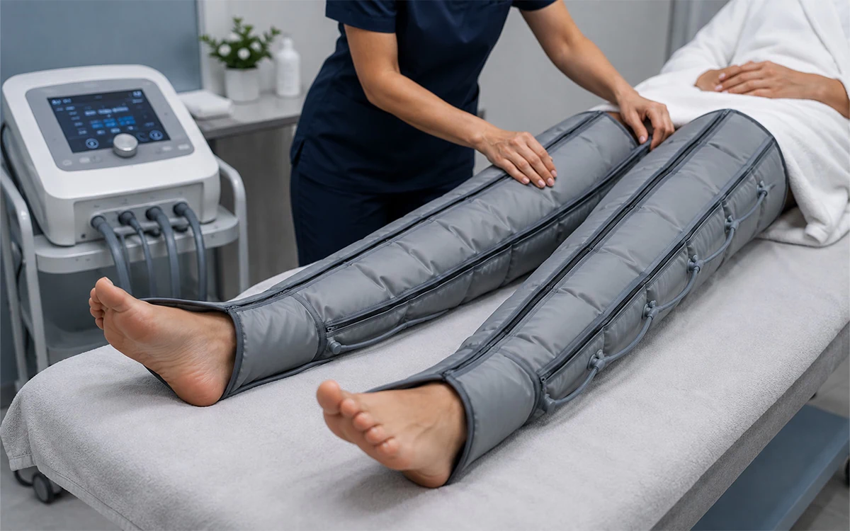
The science of compression relies on Laplace’s Law, which dictates that the pressure applied is proportional to the tension of the bandage and inversely proportional to the radius of the limb. Multi-layer compression bandages (often 3 or 4 layers) provide a "high working pressure" during walking and a "low resting pressure," which effectively pushes the pooled blood back toward the heart, reduces capillary leakage, and allows the fibrin cuffs to dissolve.
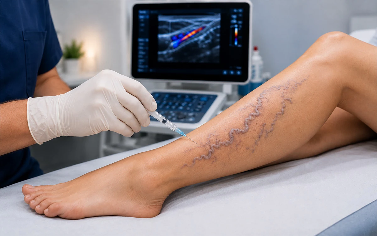
For long-term success, the "source" of the pressure must be shut off. Endovenous thermal ablation (Laser or Radiofrequency) is used to close the incompetent saphenous veins. By removing the reflux, the local venous pressure at the site of the ulcer drops significantly, which drastically reduces the time to healing and lowers the recurrence rate from nearly 30% to less than 10%.
5. The Physiological Stages of Healing
The healing of a venous ulcer occurs in four physiological stages:
- Hemostasis and Inflammation: The initial phase where the body clears debris and bacteria.
- Proliferation (Granulation): New connective tissue (granulation tissue) and microscopic blood vessels form. A healthy wound bed at this stage appears beefy red.
- Epithelialization: New skin cells move across the wound bed. This is a fragile stage where the new skin is thin and easily damaged.
- Remodeling: This stage can last for up to a year. The collagen fibers are rearranged to increase the strength of the new tissue. However, the scarred area will only ever reach about 80% of the strength of original skin.
6. Recovery and Recurrence Prevention
The "recovery" phase does not end when the skin closes. Because the underlying systemic condition (CVI) is chronic, the patient remains at high risk for recurrence. Post-ulcer management involves lifelong medical-grade compression stockings (usually Class II or III) to manage daily venous loads. Additionally, patients are educated on "skin hygiene," limb elevation, and ankle flexion exercises to maintain the efficiency of the muscle pump. Scientific management emphasizes that a "healed" ulcer is a managed condition, requiring constant vigilance to maintain tissue integrity and prevent the cycle of hypertension and ulceration from restarting.