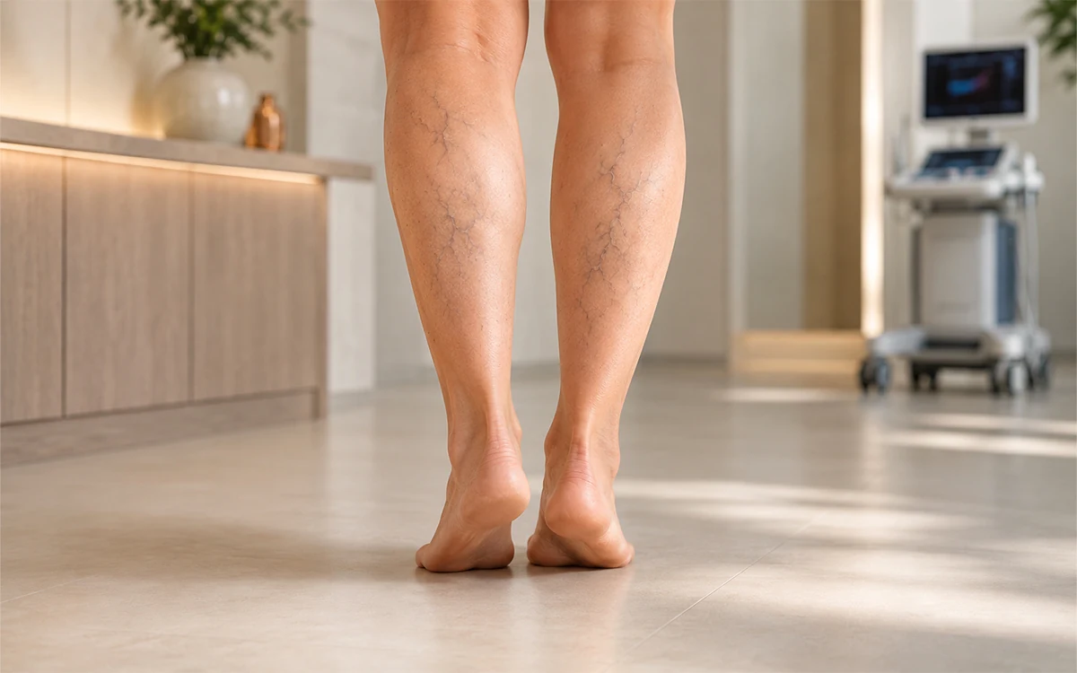
Varicose veins are a clinical manifestation of chronic venous insufficiency (CVI), characterized by dilated, tortuous, and elongated superficial veins, primarily in the lower extremities. While often perceived as a cosmetic concern, varicose veins represent a significant hemodynamic failure of the circulatory system. The scientific management of this condition has evolved from invasive surgical resection to sophisticated endovascular thermal and non-thermal ablation techniques, all aimed at correcting the underlying venous hypertension and reflux.
1. Pathophysiology and Hemodynamics
To understand the necessity of treatment, one must analyze the mechanics of venous return. In a healthy state, blood is transported from the legs to the heart against gravity via the "calf muscle pump." This system relies on one-way bicuspid valves within the veins that prevent the backward flow of blood. Varicose veins develop when these valves become incompetent—a state known as valvular reflux.
When valves fail to close properly, gravity pulls blood downward, increasing the hydrostatic pressure within the vein walls. This chronic venous hypertension leads to the secondary dilation of the vessel, which further separates the valve cusps, creating a self-perpetuating cycle of insufficiency. At a cellular level, this pressure triggers an inflammatory response in the vein wall, leading to the remodeling of the extracellular matrix, a loss of smooth muscle tone, and the characteristic protruding, twisted appearance of the vessels.
2. Clinical Indications: Who Requires Intervention?
The decision to treat varicose veins is guided by the CEAP (Clinical, Etiological, Anatomical, Pathophysiological) classification system. Scientific intervention is indicated for patients beyond the stage of simple telangiectasia who exhibit objective clinical symptoms or complications:
- Symptomatic Venous Disease (C2): Patients experience "venous claudication," which includes aching, throbbing, heaviness, and evening fatigue in the limbs. These symptoms typically worsen with prolonged standing and improve with limb elevation.
- Chronic Edema (C3): Persistent swelling of the ankle and lower leg indicates that the venous system can no longer manage the fluid volume, leading to interstitial fluid accumulation.
- Trophic Skin Changes (C4): Evidence of lipodermatosclerosis (hardening of the fat layer under the skin), hyperpigmentation (hemosiderin staining), or venous eczema. These are precursors to tissue breakdown.
- Venous Ulceration (C5–C6): Healed or active ulcers are the most severe indications, requiring immediate correction of the underlying reflux to allow the skin to heal and prevent recurrence.
- Complications: Recurrent superficial thrombophlebitis (inflammation and clotting in the superficial veins) or spontaneous hemorrhage from a friable varix.
3. Therapeutic Modalities: Scientific Execution
The primary objective of treatment is the "ablation" or removal of the truncal vein (usually the Great Saphenous Vein) where the reflux originates, thereby redirecting blood flow to the functional deep venous system.
Endovenous Thermal Ablation (EVLA and RFA)
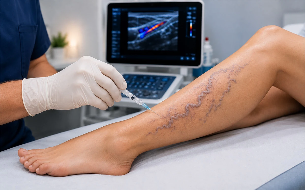
Endovenous Laser Ablation (EVLA) and Radiofrequency Ablation (RFA) are currently the gold standards of care. These procedures are performed under ultrasound guidance. A catheter is inserted into the vein and positioned just below the saphenofemoral junction. Tumescent anesthesia—a mixture of saline, lidocaine, and epinephrine—is injected around the vein. This serves three scientific purposes: it provides analgesia, acts as a heat sink to protect surrounding nerves and skin, and mechanically compresses the vein to ensure contact with the thermal probe.
The thermal energy (laser or RF) causes collagen denaturation in the vein wall, leading to immediate shrinkage and subsequent fibrotic occlusion. Over several months, the body’s inflammatory cells reabsorb the dead tissue.
Non-Thermal Ablation (Cyanoacrylate and MOCA)
For patients where thermal energy poses a risk to superficial nerves, non-thermal techniques are utilized.
- Cyanoacrylate Glue: A medical-grade adhesive is injected into the vein segments. This causes an immediate mechanical seal and a localized inflammatory reaction that leads to permanent fibrosis.
- Mechanochemical Ablation (MOCA): A rotating catheter tip physically damages the internal lining (endothelium) of the vein while simultaneously spraying a sclerosing agent. This dual action triggers rapid vessel closure without the need for heat or tumescent anesthesia.
Ultrasound-Guided Foam Sclerotherapy (UGFS)
Sclerotherapy involves the injection of a detergent-like sclerosant (such as Polidocanol). When agitated with air or CO2, it forms a foam that displaces blood and ensures maximal contact with the endothelium. This chemical injury causes the vein to collapse and eventually be replaced by a fibrous cord.
Ambulatory Phlebectomy
While the main trunk is treated with ablation, the visible bulging branches (tributaries) are often physically removed through 1–2 mm micro-incisions. This is performed using specialized hooks under local anesthesia, providing immediate relief from the physical mass of the varices.
4. The Recovery Process and Physiological Healing
The recovery period following varicose vein treatment is focused on the stabilization of the newly occluded vessel and the prevention of deep vein thrombosis (DVT).
Immediate Mobilization
Unlike historical "vein stripping" surgeries that required bed rest, modern endovascular treatment mandates immediate walking. Mobilization activates the deep vein pump, which is essential to clear any potential pro-thrombotic factors and ensure that blood flow remains vigorous in the deep system.
The Role of Compression
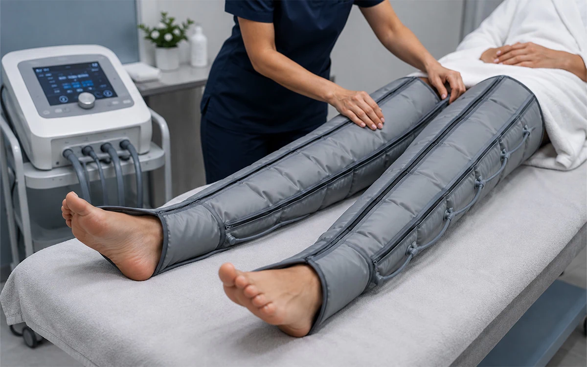
Medical compression therapy (Class II, 20–30 mmHg) is a physiological requirement post-procedure. The external pressure reduces the diameter of any patent segments, prevents the accumulation of "trapped blood" within the treated veins, and reduces post-operative edema. This ensures that the inflammatory response remains controlled and directed toward fibrosis rather than painful thrombosis.
Post-Operative Sequelae
Patients often experience a "pulling" or "tightening" sensation along the inner thigh approximately 5 to 7 days after thermal ablation. This is a positive sign of the "string sign"—the shortening of the vein as it undergoes fibrotic contraction. Bruising (ecchymosis) and mild tenderness are common as the body clears the extravasated blood and reacts to the endovascular injury.
Long-Term Resorption
The biological "disappearance" of the vein is a slow process. Macrophages and other phagocytic cells gradually break down the fibrotic remnants of the ablated vein. This process can take 3 to 6 months. During this time, follow-up Duplex Ultrasound is utilized to confirm that the treated segment remains "occluded" and that no new reflux has developed in collateral branches.
5. Maintenance and Recurrence Prevention
Varicose vein disease is a chronic condition; while the treated veins are permanently closed, the genetic and environmental factors that caused them persist. Therefore, recovery also involves lifestyle modifications. Scientific evidence suggests that maintaining a healthy Body Mass Index (BMI) and engaging in regular calf-strengthening exercises (such as swimming or cycling) reduces the load on the remaining healthy veins. Regular monitoring of the venous system allows for early identification of "neovascularization" (the growth of new, small vessels) or the progression of disease in the other limb, ensuring that venous hypertension is managed before it reaches the stage of skin breakdown or ulceration.