Thread veins, medically referred to as telangiectasias or spider veins, are small, dilated blood vessels located near the surface of the skin or mucous membranes. Typically measuring between 0.5 and 1 millimeter in diameter, these vessels appear as red, blue, or purple squiggly lines or web-like structures. While often dismissed as a purely cosmetic concern, the development of thread veins is frequently a clinical manifestation of underlying venous hypertension or localized vascular wall weakening. The scientific management of this condition involves precise diagnostic mapping and the application of chemical or thermal energy to induce vessel fibrosis and resorption.
1. Pathophysiology and Etiology: Why Treatment Is Sought
The formation of thread veins is driven by the dilation of pre-existing small vessels. In the lower extremities, this is often the result of "micro-reflux." Although the large valves in the deep venous system may be functioning correctly, the smaller valves in the reticular veins (the green-tinted veins just below the skin) may fail. This failure increases the hydrostatic pressure within the smallest capillaries, causing them to distend and become visible.
Several factors contribute to this process:
- Genetic Predisposition: A primary factor is an inherited weakness in the vessel walls or valve structures.
- Hormonal Influence: Estrogen is known to relax the smooth muscle in vein walls, making them more susceptible to dilation. This explains the increased prevalence during pregnancy, menopause, or with the use of oral contraceptives.
- Environmental and Lifestyle Factors: Prolonged standing or sitting increases venous pressure. Additionally, ultraviolet (UV) radiation can damage the collagen supporting the vessel walls, particularly on the face, leading to permanent dilation.
- Inflammatory Skin Conditions: Conditions such as rosacea can cause chronic vasodilation, eventually leading to permanent telangiectasias.
From a clinical perspective, treatment is indicated not only for the psychological impact of the condition but also to alleviate physical symptoms such as localized itching, burning sensations, or a feeling of "heaviness" in the affected area. Furthermore, treating thread veins can prevent the progression of localized venous insufficiency.
2. Diagnostic Necessity: The Role of Duplex Ultrasound
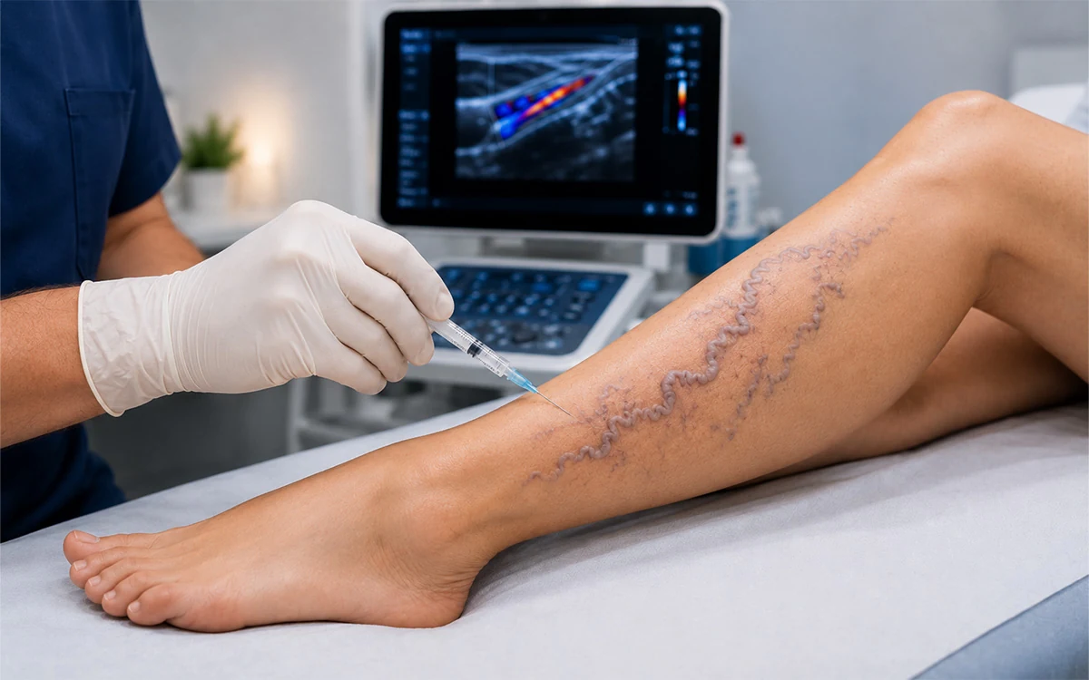
Before any treatment is initiated, a scientific assessment is mandatory. The "gold standard" for diagnosis is the Color Duplex Ultrasound. This is used to rule out underlying "feeder veins" or saphenous vein reflux. If a clinician treats surface thread veins while a deeper source of reflux (venous hypertension) remains unaddressed, the treatment is highly likely to fail, leading to rapid recurrence or a phenomenon known as "telangiectatic matting," where a dense forest of tiny new vessels forms in response to the untreated pressure.
3. Primary Treatment Modalities
The goal of treatment is the permanent destruction of the vessel lumen. This is achieved through two primary scientific methods: Microsclerotherapy and light-based therapies.
Microsclerotherapy
Microsclerotherapy is the clinical gold standard for thread veins on the legs. It involves the injection of a sclerosing agent (a liquid chemical) directly into the target vessel using an ultra-fine needle. Common agents include Polidocanol or Sodium Tetradecyl Sulfate.
The mechanism of action is a controlled chemical endovenous injury. The sclerosant displaces the blood and reacts with the endothelial lining of the vessel wall. This causes "endothelial denudation"—the stripping away of the inner cell layer—triggering a localized inflammatory cascade. This leads to the formation of a small, controlled thrombus (clot) and subsequent fibrosis. Over time, the vessel turns into a cord of connective tissue that the body’s macrophages eventually break down and reabsorb.
Selective Photothermolysis (Laser and IPL)
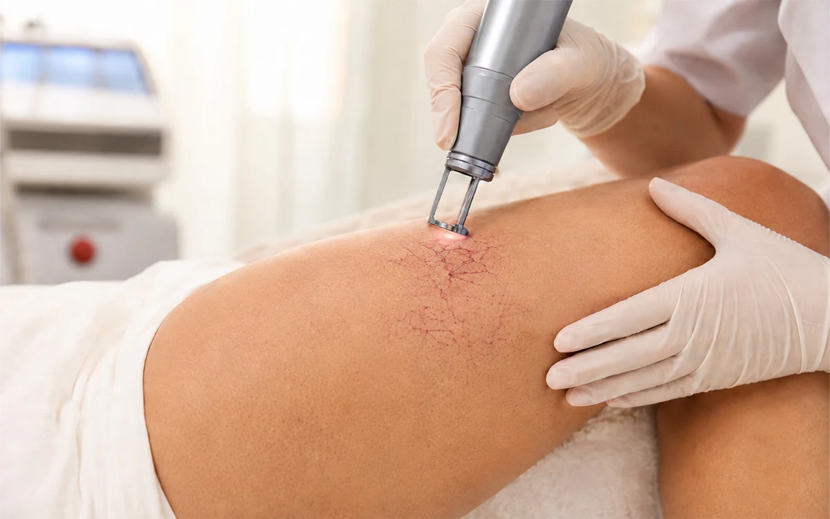
For very fine thread veins, particularly those on the face where needles are less practical, light-based treatments are utilized. This relies on the principle of selective photothermolysis.
A specific wavelength of light (often the 1064 nm Nd:YAG laser or Intense Pulsed Light) is directed at the skin. The light energy is selectively absorbed by the chromophore "hemoglobin" within the red blood cells. The energy is converted into heat, which is then transferred to the vessel wall. The temperature rises sufficiently to cause thermal damage to the vessel (coagulation) without damaging the surrounding skin tissue. The damaged vessel then collapses and is removed by the body’s natural healing processes.
4. The Recovery Process and Physiological Adaptation
Recovery from thread vein treatment is a biological process that occurs over weeks or months, rather than days. It is important to understand that the appearance often worsens before it improves.
The Inflammatory Phase (Weeks 1–3)
Immediately following sclerotherapy, the treated veins may appear darker, bruised, or "trapped." This is a sign that the sclerosant has worked; the blood inside has coagulated, and the inflammatory process of vessel closure has begun. Patients may feel slight firmness along the line of the vein.
Compression Therapy
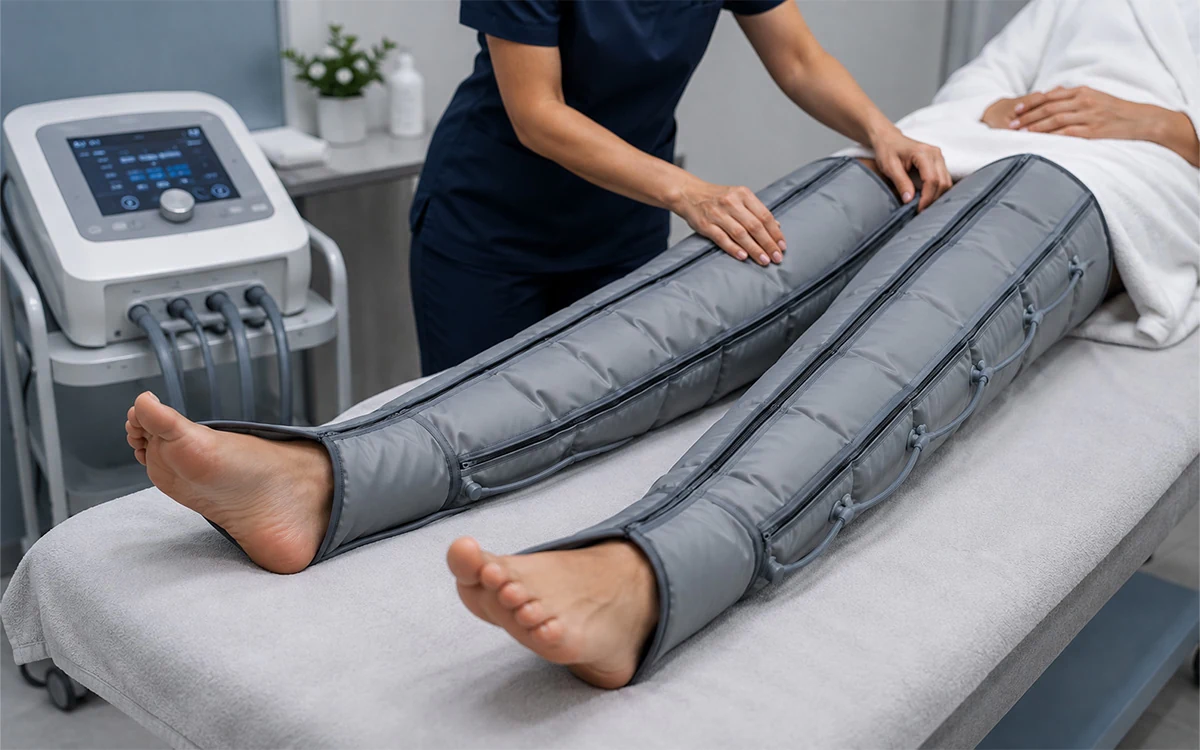
The use of medical-grade compression stockings is scientifically vital following leg treatments. Compression reduces the diameter of the treated vessels, ensuring that the walls remain in contact for successful fibrosis and minimizing the amount of trapped blood. It also helps lower the local venous pressure, which speeds up the healing of the micro-punctures.
Hemosiderin Staining and Resolution
As the body breaks down the treated vein, iron from the blood (hemosiderin) can occasionally leave a temporary brownish stain on the skin. This is not a scar but a pigmentary change that the body typically clears over 3 to 12 months.
Cellular Resorption
The final stage of recovery involves the lymphatic system and macrophages. Once the vessel has been fibrosed (turned to scar tissue), the body recognizes it as waste. It is slowly digested and removed. For this reason, the final results of a single treatment session are often not evaluated for at least 8 to 12 weeks.
5. Long-Term Maintenance and Prevention
While the treated veins are permanently destroyed, the underlying biological tendency to form new thread veins remains. Therefore, management often involves a long-term strategy. This includes UV protection to prevent facial telangiectasias and the management of venous health through exercise and weight control for the legs. Regular follow-up appointments allow clinicians to identify new "feeder veins" early, maintaining the integrity of the peripheral vascular system and preventing the recurrence of visible symptoms. Through this scientific, staged approach, the physiological and symptomatic aspects of thread vein disease can be effectively controlled.