
Chronic Venous Disease (CVD) represents a spectrum of morphological and functional abnormalities of the venous system, primarily affecting the lower extremities. It is a progressive condition that, if left unaddressed, can lead to significant morbidity. Understanding the scientific basis of vein treatment requires an exploration of venous hemodynamics, the technological evolution of interventional procedures, and the physiological requirements of post-operative care.
1. The Pathophysiology of Venous Insufficiency
To understand why treatment is necessary, one must first understand the “muscle pump” mechanism. Unlike the arterial system, which relies on the heart’s pumping action, the venous system in the legs must transport blood against gravity. This is achieved through the contraction of calf muscles and the presence of bicuspid valves that prevent retrograde flow (reflux).
Venous disease occurs when these valves become incompetent or when the vein walls lose their elasticity (dilatation). This failure leads to venous reflux, where blood pools in the lower limbs, causing an increase in intravenous pressure—a state known as chronic venous hypertension. This high pressure triggers a cascade of inflammatory responses, leading to capillary leakage, tissue edema, and eventually, skin changes or ulceration.
2. Clinical Indications: Who Requires Treatment?
Treatment is not merely an aesthetic choice; it is a clinical necessity for individuals classified under the CEAP (Clinical, Etiological, Anatomical, Pathophysiological) classification system. Scientific intervention is typically indicated for:
- Symptomatic Patients: Those experiencing “venous claudication,” characterized by heavy limbs, aching, throbbing, and nocturnal muscle cramps that intensify at the end of the day.
- Chronic Edema: Patients with persistent swelling that indicates the lymphatic system is overwhelmed by venous fluid extravasation.
- Trophic Skin Changes: Individuals showing signs of lipodermatosclerosis (thickening of the subcutaneous tissue), hyperpigmentation (hemosiderin staining from burst capillaries), or venous eczema.
- Venous Ulceration (C5–C6): Patients with active or healed ulcers require urgent intervention to correct the underlying reflux and allow for tissue healing.
- Thrombotic Risk Management: Those with a history of superficial thrombophlebitis or at risk of deep vein thrombosis (DVT) due to stasis.
3. Modern Therapeutic Modalities
The goal of modern treatment is the “ablation” or removal of the diseased vessel, redirecting blood flow to healthy, functional veins.
A. Endovenous Thermal Ablation (EVLA and RFA)
Endovenous Laser Ablation (EVLA) and Radiofrequency Ablation (RFA) are the gold standards for treating the Great Saphenous Vein (GSV). Under ultrasound guidance, a catheter is inserted into the vein. Thermal energy is applied to the vein wall, causing collagen denaturation and immediate contraction. This process leads to fibrotic occlusion—the vein is essentially turned into scar tissue, which the body eventually reabsorbs.
B. Sclerotherapy and Ultrasound-Guided Foam Sclerotherapy (UGFS)
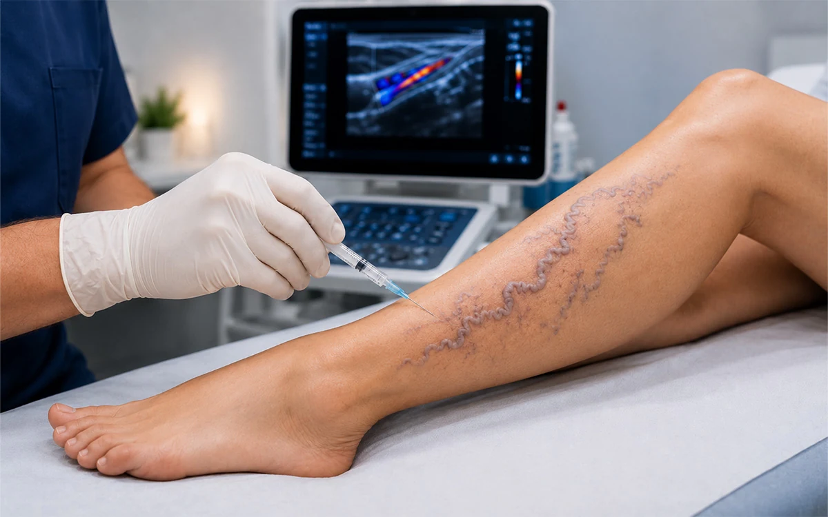
This involves the injection of a sclerosing agent (such as Polidocanol or Sodium Tetradecyl Sulfate). The chemical irritates the endothelial lining of the vessel, causing a localized inflammatory reaction that leads to the vessel's closure. Foam sclerotherapy is particularly effective for tortuous (twisted) veins where a straight catheter cannot pass.
C. Non-Thermal Non-Tumescent (NTNT) Techniques
Recent scientific advancements have introduced medical adhesives (Cyanoacrylate glue). This technique does not require heat, eliminating the risk of nerve injury and removing the need for “tumescent anesthesia” (large volumes of fluid injected around the vein). The glue polymerizes upon contact with blood, mechanically sealing the vein instantly.
D. Microphlebectomy
For large, bulging surface varicosities, a microphlebectomy is performed. Through 1–2 mm micro-incisions, the clinician physically removes segments of the vein. Because the incisions are so small, they typically heal without sutures and with minimal scarring.
4. The Recovery Process and Physiological Adaptation
Post-treatment recovery is a critical phase where the body adapts to new hemodynamic patterns.
Immediate Post-Operative Phase (0–48 Hours)
The primary goal is the prevention of DVT. Unlike traditional “stripping” surgery, modern ablation requires immediate mobilization. Walking encourages the calf muscle pump to move blood through the deep venous system, reducing stasis. Patients may experience mild bruising or a “tugging” sensation along the treated vein as the inflammatory process of fibrosis begins.
The Role of Compression Therapy
Medical-grade compression stockings (usually 20–30 mmHg) are scientifically vital. They provide external hydrostatic pressure that counteracts internal venous pressure, minimizes edema, and ensures the walls of the ablated vein remain in contact during the early stages of fibrosis.
Cellular Absorption (Weeks to Months)
The “closed” vein is no longer a functional vessel but a cord of connective tissue. Over several months, macrophages (immune cells) gradually break down this tissue. During this time, the skin hyperpigmentation often fades as the iron deposits (hemosiderin) from stagnant blood are cleared by the lymphatic system.
5. Why Treatment Cannot Be Ignored: The Risks of Neglect
If the underlying venous hypertension is not corrected, the disease follows a predictable degenerative path. The most severe complication is venous stasis ulceration. These ulcers occur because the high venous pressure prevents fresh, oxygenated arterial blood from reaching the skin cells, leading to localized tissue death (necrosis). Furthermore, stagnant blood in dilated veins is prone to spontaneous clotting, which can migrate to the deep veins, causing a life-threatening pulmonary embolism.
6. Conservative Management and Prevention
While interventional procedures fix the damaged valves, venous health is maintained through lifestyle physiology:
- Venoactive Pharmacotherapy: Certain flavonoids can improve venous tone and reduce capillary permeability.
- Weight Regulation: Reducing intra-abdominal pressure lowers the resistance against which leg blood must travel.
- Kinesiotherapy: Strengthening the gastrocnemius (calf) muscle enhances the efficiency of the venous return.
Conclusion
Venous disease treatment has transitioned from invasive surgical resection to sophisticated, image-guided endovascular medicine. By addressing the root cause—valvular reflux and venous hypertension—these treatments restore normal circulatory hemodynamics. For the patient, this translates to the resolution of pain, the prevention of skin breakdown, and a significant reduction in the risk of thromboembolic events. Success depends on a combination of precise technological intervention and the physiological support of the recovery process through compression and movement.